Digital Twin for PET Scanning
PhD Project by Christian Hinge
The aim of this project is to enhance the diagnostic potential of whole-body Positron Emission Tomography/Computed Tomography (PET/CT) imaging by creating personalized synthetic healthy PET baselines using advanced Deep Learning techniques.
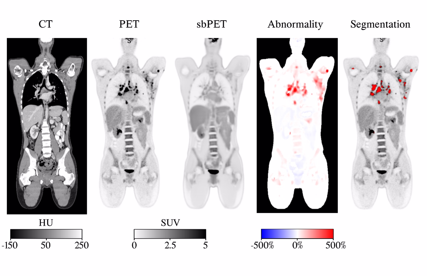
Project BackgroundWhole-body PET/CT imaging with FDG tracers is an invaluable diagnostic tool widely used in hospitals for detecting, diagnosing, and monitoring various diseases. However, standard analysis methods lack personalized healthy control images, reducing their precision and limiting the full diagnostic and prognostic potential of PET/CT imaging. This project addresses this limitation by introducing advanced Deep Learning techniques to synthesize personalized PET images based on the patient's own whole-body CT scans. This approach enables the development of images that reflect the individual patient's anatomical and physiological characteristics, increasing the accuracy of differentiating between normal and diseased states.
Project PotentialThe project has the potential to detect deviations and subtle changes in organ uptake patterns by comparing the patient's digital twin with the actual PET scan. For example, in diabetes, a personalized healthy PET image can assess the disease by comparing it to the actual observed uptake. This project represents an innovative approach to improving diagnostic analysis methods, enhancing the accuracy of diagnoses, and increasing the effectiveness of treatment for individual patients.

Figure 1: Application of healthy PET for lymphoma detection. From left: CT, PET, sbPET: Synthetic healthy PET image, Abnormality: difference between PET and synthetic PET, Segmentation: identified cancerous areas.