Quantitative Image-Derived Input Function for the Estimation of Cerebral Blood Flow
Project by Thomas Lund Anderesen
The quantification of cerebral blood flow using [15O]H2O PET relies on precise input function estimation, conventionally obtained via arterial blood sampling. This study investigates an automated approach to image-derived input function (IDIF) extraction using a long axial field-of-view (LAFOV) PET/CT scanner, with the aim of replacing invasive procedures.
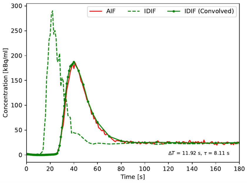
Project BackgroundThe LAFOV PET/CT scanner enables a direct and non-invasive sampling of the blood activity concentration by extracting the arterial input function (AIF) directly from the PET image data. In this project the extracted IDIF was compared with the AIF obtained through simultaneous arterial blood sampling. Modelling of regional cerebral blood flow (rCBF) using kinetic compartment modelling with [15O]H2O PET data using IDIF and AIF was compared using the two blood input functions.
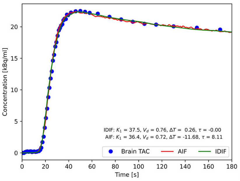
Project ImplementationThe IDIF closely matched the gold standard across multiple metrics, including peak shape and area under the curve.
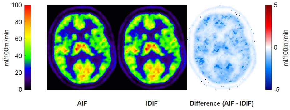
Cerebral blood flow estimates based on IDIF offered a reliable alternative to invasive blood sampling, as the input function can be measured directly from the PET image data. The voxel-wise perfusion maps (Figure 3) with quantitative differences between AIF- and IDIF-based estimates were minimal across the brain. Ultimately, the extraction of the blood concentration function has been implemented clinically for the benefit for patients.