Device-Less Data-Driven Cardiac and Respiratory Gating in PET Imaging
Project by Nanna Overbeck
Respiratory and cardiac motion during Positron Emission Tomography (PET) scans can cause image blurring, reducing diagnostic accuracy and affecting treatment planning. This project introduces a device-less, data-driven approach utilizing high-sensitivity long axial field-of-view (LAFOV) PET histo images. By enabling gated image reconstruction, the method improves image clarity, enhances tumor detectability, and provides more reliable diagnostic information without the need for external devices.
Project BackgroundPET, combined with Computed Tomography (CT), is a valuable tool for diagnosing and treatment planning in oncology, neurology, and cardiology. However, motion during PET scans can lead to reduced image quality, which may lead to inaccurate assessments of tumor activity, size, and visibility.
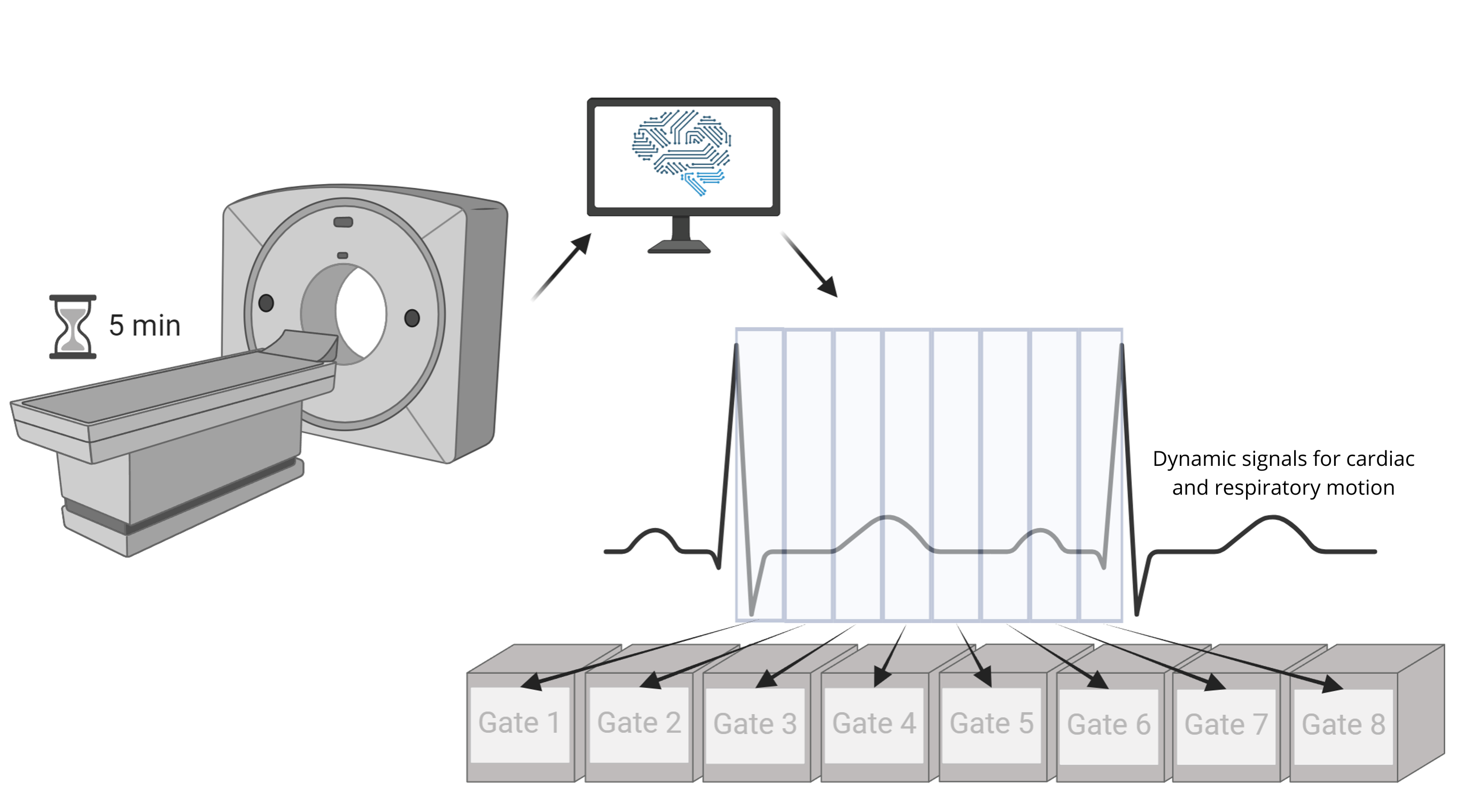
Gated image reconstruction can address these issues by dividing scan data into phases of the motion cycle. Traditional gating methods rely on external devices, which increase preparation time and complexity.
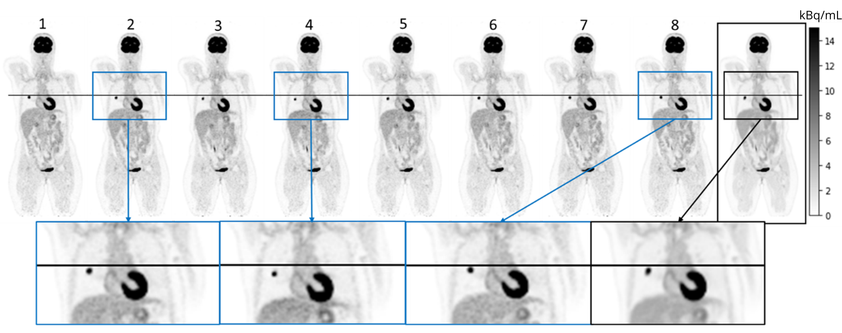
This project explores a device-less alternative using LAFOV [18F]FDG PET histo images and Fourier Transform (FT) analysis to estimate cardiac and respiratory motion. Results from 18 patients show that this method reliably matches reference measurements. Gated reconstructions demonstrated increased tumor maximum standardized uptake values (SUVmax) and reduced tumor volume, improving tumor quantification and overall diagnostic precision.
Project PotentialThis device-less, data-driven method has the potential to optimize PET imaging by reducing preparation time and eliminating the need for external devices. It enhances motion correction, leading to more accurate tumor measurements and improved diagnostic accuracy. LAFOV PET histo images have demonstrated consistent and reliable motion estimation, with gated reconstructions effectively isolating heart and lung motion. This approach enhances image clarity, making it particularly valuable for motion-sensitive cases such as lung and cardiac imaging, with potential for clinical integration.
Gated PET Videos of Cardiac and Respiratory Motion
Approach: Data-Driven gated PET reconstruction
Results: Data-Driven gated PET reconstruction